
Did you know that the average animal cell is only about 10 to 30 micrometers in size? That’s smaller than the width of a human hair!
As you delve into the fascinating world of animal cell biology, you will uncover the intricate structure, parts, and functions that allow these tiny powerhouses to carry out the essential processes of life.
From the cell membrane that controls what enters and exits the cell, to the nucleus that holds the genetic material, each component plays a vital role in the overall functioning of the cell.
But that’s not all – there are numerous organelles and structures within the cell that contribute to its complexity and efficiency.
So, get ready to explore the inner workings of animal cells and discover the remarkable building blocks that make life possible.
Animal Cell Size and Shape
Animal cell size and shape vary greatly, with the largest animal cell being the ostrich egg and the smallest being neurons. Animal cells come in various shapes, and they lack a cell wall, allowing for diverse shapes. Generally, animal cells are irregular in shape. The size and shape of an animal cell are determined by its function and the specific organs it contains.
The largest animal cell, the ostrich egg, is visible to the naked eye and measures about 5 inches in diameter. In contrast, neurons, the smallest animal cells, are microscopic and have long, thin extensions called axons and dendrites.
The irregular shape of animal cells allows them to fit together tightly, forming tissues and organs. This shape enables cells to communicate and interact with one another. The plasma membrane, which surrounds animal cells, plays a crucial role in maintaining the shape and integrity of the cell.
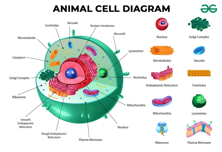
Animal Cell Organelles
Now let’s explore the fascinating world of animal cell organelles. These structures play a crucial role in the structure and function of the cell.
Key organelles such as the nucleus, cytoplasm, Golgi apparatus, lysosomes, and microtubules are involved in vital cellular processes such as DNA regulation, protein synthesis, cell metabolism, and cell division.
Understanding the functions of these organelles will give you a deeper insight into the complexity of animal cells.
Structure and Function
The organelles found within animal cells perform vital functions that contribute to the overall structure and function of the cell.
The nucleus, surrounded by a double-layered nuclear membrane, holds genetic material in the form of DNA and controls and regulates cell activities.
The cytoplasm, a gel-like material, contains various organelles, including mitochondria, which play a role in cell metabolism.
The Golgi apparatus is responsible for transporting, modifying, and packing proteins and lipids.
Lysosomes function in digestion, excretion, and cell renewal by breaking down macromolecules.
The cytoskeleton, made up of proteins, organizes cell components and maintains cell shape.
Microtubules, long hollow cylinders, assist in transportation, provide structural support, and make up cilia and flagella.
Centrioles are involved in cell division, while peroxisomes are involved in lipid metabolism and detoxification.
Cilia and flagella are locomotive projections, and endosomes are involved in folding the plasma membrane.
Vacuoles store various substances, and microvilli increase surface area for absorption.
Key Organelles
One of the key organelles found in animal cells is the nucleus, which plays a crucial role in controlling and regulating cell activities through the storage of genetic material in the form of DNA. The nucleus is a spherical organelle surrounded by a double-layered nuclear membrane. It holds the genetic material and controls cell activities. Transcription, the process of synthesizing RNA from DNA, occurs within the nucleus.
Another important organelle in animal cells is the cytoplasm, a gel-like substance that houses various organelles. Mitochondria, which are membrane-bound organelles, are found within the cytoplasm and play a significant role in cellular metabolism.
Together, the nucleus and cytoplasm form the core of animal cell function, ensuring proper cellular processes and maintaining cell integrity.
Cellular Metabolism
Animal cell organelles play a crucial role in cellular metabolism by facilitating various biochemical processes and reactions.
The mitochondria, for example, are responsible for producing energy in the form of ATP through cellular respiration. They break down glucose and other molecules to generate ATP, which powers cellular activities.
The endoplasmic reticulum (ER) is involved in protein synthesis, folding, and modification. It also plays a role in lipid metabolism and detoxification.
The Golgi apparatus modifies, sorts, and packages proteins and lipids for transport to their final destinations.
Lysosomes contain enzymes that break down macromolecules for digestion and recycling.
Plasma Membrane (Cell Membrane)
The plasma membrane, also known as the cell membrane, serves as a barrier and regulator for animal cells. It controls the movement of molecules in and out of the cell, allowing for selective permeability.
Additionally, the proteins and lipids in the plasma membrane play a crucial role in cell communication and protection.
Barrier and Regulation
The plasma membrane, also known as the cell membrane, serves as a thin, semi-permeable barrier that encloses and protects the contents of an animal cell. It regulates the molecules that pass into and out of the cell, ensuring that essential substances are allowed in while harmful substances are kept out.
The plasma membrane consists of a lipid bilayer embedded with proteins and carbohydrates. These proteins and lipids play a crucial role in cell communication and recognition.
The plasma membrane also maintains the cell’s shape and integrity by providing structural support. It’s essential for maintaining homeostasis within the cell, allowing it to function properly and respond to changes in its environment.
Cell Communication and Protection
To understand how animal cells communicate and protect themselves, we must now focus on the plasma membrane, the thin barrier that regulates the flow of molecules in and out of the cell.
The plasma membrane plays a crucial role in cell communication and protection. It encloses and protects the cell content, while also allowing selective transport of molecules.
Proteins and lipids embedded in the plasma membrane facilitate cell communication by transmitting signals between cells. These signals can regulate various cellular processes, such as growth, differentiation, and immune responses.
Additionally, the plasma membrane acts as a protective barrier, preventing harmful substances from entering the cell and maintaining the internal environment necessary for cell function.
Nucleus
Surrounded by a double-layered nuclear membrane, the nucleus of an animal cell plays a crucial role in controlling and regulating cellular activities. It’s a spherical organelle that holds genetic material in the form of DNA.
The nucleus is responsible for controlling the activities of the cell by directing the synthesis of proteins and other molecules through a process called transcription. Within the nucleus, DNA is organized into structures called chromosomes.
The nucleus also contains a dense region called the nucleolus, which is involved in the production of ribosomes. The nuclear membrane has pores that allow for the movement of molecules in and out of the nucleus. These pores are important for the transport of RNA molecules, proteins, and other molecules that are necessary for cellular processes.
Cytoplasm
Now let’s talk about the cytoplasm, the gel-like material in animal cells that houses various organelles.
It’s where you can find important cellular components and plays a crucial role in cell metabolism.
Mitochondria, which are responsible for energy production, are also found in the cytoplasm.
Gel-Like Cell Material
The cytoplasm in animal cells is a gel-like substance that houses various organelles and plays a crucial role in cell metabolism. It’s a semi-fluid material that fills the space between the cell membrane and the nucleus.
The cytoplasm contains numerous organelles, such as mitochondria, which produce energy for the cell, and ribosomes, which are involved in protein synthesis.
Additionally, the cytoplasm provides a medium for chemical reactions to occur, serving as a site for metabolic processes. It also aids in the transport of molecules within the cell, allowing for the movement of organelles and the distribution of nutrients and waste products.
Organelles in Cytoplasm
As we shift our focus to the organelles within the cytoplasm, let’s explore the intricate structures that contribute to the functionality of animal cells.
The cytoplasm is a gel-like material that contains various organelles essential for cellular processes. One of the prominent organelles found in the cytoplasm is the mitochondria, which are responsible for generating energy through cellular respiration.
Additionally, the cytoplasm houses important cellular components such as the ribosomes, which are involved in protein synthesis. It also plays a crucial role in cell metabolism, providing a medium for biochemical reactions to occur.
The cytoplasm acts as a supportive environment for other organelles and is vital for the overall functioning of the animal cell.
Golgi Apparatus
Located within the cytoplasm of animal cells, the Golgi apparatus is a crucial organelle responsible for the transport, modification, and packaging of proteins and lipids. It consists of a stack of flattened, membrane-bound sacs called cisternae.
The Golgi apparatus receives proteins and lipids from the endoplasmic reticulum and modifies them through various processes, such as glycosylation, where sugar molecules are attached to proteins. It also plays a role in sorting and packaging these molecules into vesicles for transport to their final destinations.
The Golgi apparatus is divided into distinct regions, including the cis, medial, and trans compartments. Each compartment has specific enzymes that carry out different modifications to the proteins and lipids passing through.
Additionally, the Golgi apparatus is involved in the production of lysosomes, which are organelles responsible for the digestion of cellular waste materials.
Lysosomes
Lysosomes, the cellular recycling centers, break down macromolecules into smaller molecules for digestion, excretion, and cell renewal. These round subcellular organelles have a membrane and contain lysosomal enzymes that play a crucial role in cellular processes.
Lysosomes are involved in the digestion of cell nutrients, breaking them down into smaller molecules that can be used by the cell. They also aid in the excretion of waste materials from the cell. Additionally, lysosomes are responsible for cell renewal by breaking down cellular components that are no longer needed, allowing for the recycling of valuable materials.
Lysosomal enzymes are specialized proteins that catalyze the breakdown of various macromolecules, including proteins, lipids, carbohydrates, and nucleic acids. This process, known as hydrolysis, occurs within the acidic environment of the lysosome.
Lysosomes are essential for maintaining cellular homeostasis and ensuring the proper functioning of animal cells.
Cytoskeleton
The cytoskeleton, a complex network of proteins, provides structural support and organizes cell components in animal cells. It’s made up of three main components: actin filaments, microtubules, and intermediate filaments.
Actin filaments play a crucial role in maintaining cell shape, mediating cell activities, and assisting in mitotic cell division. They provide structural support and help the cell maintain its shape.
Microtubules, on the other hand, assist in mitosis, provide structural support, and make up important cell structures such as cilia and flagella. They’re long, hollow cylinders made of tubulin protein and are involved in cell locomotion and transportation of organelles.
Intermediate filaments provide mechanical strength to the cell, helping it withstand mechanical stress. They’re important for maintaining cell shape and integrity.
Microtubules
Microtubules, composed of tubulin protein, are essential components of the cytoskeleton in animal cells. These long, hollow cylinders are found throughout the cytoplasm and play crucial roles in various cellular processes.
One important function of microtubules is the transportation of organelles within the cell. They serve as tracks along which motor proteins move, facilitating the movement of vesicles, mitochondria, and other cellular components.
Microtubules also provide structural support to the cell, helping to maintain its shape and integrity.
During cell division, microtubules form spindle fibers that are responsible for separating chromosomes. They attach to the centromeres of chromosomes and ensure their proper alignment and distribution to daughter cells.
Microtubules are also the main components of cilia and flagella, which are locomotive projections found on the cell surface. In cilia, microtubules are organized in a ‘9+2’ arrangement, while in flagella, they’re arranged in a ‘9+0’ pattern. Through the coordinated movement of these microtubules, cilia and flagella enable the cell to move fluids and particles or aid in the locomotion of sperm cells.
Centrioles
Centrioles, essential organelles in animal cells, play a crucial role in cell division and the organization of microtubules. These cylindrical structures are composed of nine microtubule bundles arranged in triplets. They’re found in pairs called centrosomes and are located near the nucleus.
During cell division, centrioles replicate, resulting in the formation of two centrosomes. Centrioles are involved in the formation of the mitotic spindle, which is responsible for separating chromosomes during cell division. They also help in the organization of microtubules, which form the cytoskeleton of the cell and provide structural support.
Additionally, centrioles are involved in the movement of organelles within the cell. Through their anchoring function, they serve as attachment sites for microtubules, which aid in the transportation of substances.
Peroxisomes
Let’s now explore the functions and structure of peroxisomes in animal cells.
Peroxisomes are tiny bodies found in the cytoplasm, bound by a membrane, and involved in lipid metabolism. They’re the most common micro-bodies in the cell cytoplasm and play a crucial role in detoxifying chemicals by producing hydrogen peroxide.
Peroxisome Functions
Peroxisomes, small membrane-bound organelles found in the cytoplasm of animal cells, play a crucial role in lipid metabolism and the detoxification of chemicals by producing hydrogen peroxide.
These organelles contain enzymes that break down fatty acids and convert them into a form that can be used for energy production. Additionally, peroxisomes are involved in the synthesis of certain lipids, such as cholesterol and plasmalogens.
They also play a role in the detoxification of harmful substances by breaking them down into less toxic compounds. The production of hydrogen peroxide within peroxisomes is a key step in these detoxification processes.
Peroxisome Structure
The structure of peroxisomes, small membrane-bound organelles found in the cytoplasm of animal cells, plays a crucial role in their functions.
Peroxisomes are spherically shaped and are bound by a membrane. They’re the most common micro-bodies in the cell cytoplasm.
Peroxisomes are involved in lipid metabolism and detoxify chemicals by producing hydrogen peroxide. They contain enzymes that break down fatty acids and convert them into molecules that can be used by the cell for energy.
Additionally, peroxisomes are responsible for the breakdown of toxic substances, such as ethanol, and the synthesis of important molecules, including cholesterol and bile acids.
The structure of peroxisomes allows them to carry out these vital functions efficiently and effectively.
Cilia and Flagella
Cilia and flagella, specialized locomotive projections found on the surface of animal cells, play crucial roles in cell movement and fluid propulsion.
Cilia are short, hair-like structures that cover the cell surface and work together in a coordinated motion to move fluids and particles across the cell. They’re found in the respiratory tract, where they help to move mucus and trapped particles out of the lungs.
Flagella, on the other hand, are longer and less numerous than cilia. They act as whip-like structures and are responsible for cell movement. One well-known example of flagella is found in sperm cells, where they enable the sperm to swim towards the egg for fertilization.
Both cilia and flagella are made up of strands of filaments called microtubules. These microtubules are arranged in a specific pattern and are connected by motor proteins called dynein. The movement of dynein along the microtubules causes the cilia and flagella to bend, generating a wave-like motion that propels the cell or moves fluid around the cell.
Endosomes
Endosomes play a crucial role in the folding of the plasma membrane and the removal of waste materials from the cell through endocytic processes. These membranous organelles are formed by endocytosis and are found in the cell cytoplasm. Endosomes are involved in the internalization of extracellular substances, such as nutrients and signaling molecules, through receptor-mediated endocytosis. This process allows the cell to regulate its nutrient intake and respond to external signals.
Once inside the cell, endosomes undergo a series of maturation steps. They fuse with lysosomes, which contain digestive enzymes, to form endolysosomes. These endolysosomes facilitate the degradation of internalized materials, including proteins, lipids, and carbohydrates. The breakdown products are then released into the cytoplasm for further processing or excretion.
In addition to their role in endocytosis, endosomes are also involved in the recycling of membrane proteins. They sort and transport internalized receptors back to the plasma membrane or to other cellular compartments for reuse or degradation. This process is important for maintaining the integrity and functionality of the plasma membrane.
Vacuoles
What is the role of vacuoles in animal cells?
Vacuoles are fluid-filled organelles enclosed by a membrane, known as the tonoplast. They’re membrane-bound sacs found in the cytoplasm of animal cells.
The primary function of vacuoles is to store various substances such as food, water, carbohydrates, and waste materials. They can also remove toxic substances and poorly folded proteins from the cell.
Vacuoles play a crucial role in maintaining cellular homeostasis by regulating the inflow and outflow of small molecules through the tonoplast. Additionally, they contribute to the structural integrity of the cell by exerting turgor pressure, which helps maintain the shape of the cell.
The size and number of vacuoles can vary depending on the specific cell type and its function. For example, plant cells typically have larger central vacuoles that store water, while animal cells may have multiple smaller vacuoles scattered throughout the cytoplasm.
Microvilli
Microvilli are surface protrusions that increase the surface area of certain cells, allowing for enhanced absorption and anchoring functions. These tiny finger-like projections can be found in the intestinal lining, on the surface of egg cells, and on white blood cells. Microvilli are formed from accessory proteins of actin filaments.
In the small intestines, microvilli increase the surface area, allowing for more efficient absorption of nutrients. Their presence greatly enhances the ability of the intestines to absorb nutrients from digested food, as the increased surface area provides more sites for absorption to occur.
In the case of egg cells, microvilli play a crucial role in fertilization. They anchor sperm cells to the egg surface, facilitating the process of fertilization. Without microvilli, the sperm wouldn’t be able to properly attach to the egg, hindering the fertilization process.
Microvilli also serve as anchors for white blood cells in the circulatory system. They help white blood cells adhere to the walls of blood vessels, allowing them to migrate to sites of infection or injury. This is essential for the body’s immune response.
Conclusion
The intricate structure and functions of animal cells are vital for the proper functioning of living organisms.
From the plasma membrane that regulates the flow of substances in and out of the cell, to the nucleus that controls cell activities, and the various organelles each contributing to specific cell processes, animal cells are remarkable building blocks of life.
Understanding their structure and functions is crucial for gaining insights into the complex world of biology.

Erzsebet Frey (Eli Frey) is an ecologist and online entrepreneur with a Master of Science in Ecology from the University of Belgrade. Originally from Serbia, she has lived in Sri Lanka since 2017. Eli has worked internationally in countries like Oman, Brazil, Germany, and Sri Lanka. In 2018, she expanded into SEO and blogging, completing courses from UC Davis and Edinburgh. Eli has founded multiple websites focused on biology, ecology, environmental science, sustainable and simple living, and outdoor activities. She enjoys creating nature and simple living videos on YouTube and participates in speleology, diving, and hiking.