

Are you ready to embark on a journey of discovery, as we shine a light on the fascinating world that can be unveiled through a light microscope?
Like a window into a hidden realm, this powerful tool harnesses the magic of visible light to detect and magnify small objects. Through the lens of refraction and the intricate design of glass lenses, we can explore the microscopic wonders that lie beyond our naked eye.
But that’s just the beginning. There are various types of light microscopes, each with its own specialized applications and functionalities, waiting to be explored. From the brightfield microscope, used to study microbial cultures, to the phase contrast microscope, ideal for observing transparent specimens, and the dark-field and fluorescent microscopes with their unique capabilities in medical diagnostics and research.
But hold on, we’re not done yet. We will also uncover the intricate parts that make up a light microscope, allowing it to reveal the hidden secrets of the microscopic world.
So, are you ready to embark on this enlightening journey with us?
Definition of a Light Microscope
A light microscope is a scientific instrument that utilizes visible light to detect and magnify small objects, allowing for detailed examination and analysis. It’s a vital tool in various fields such as biology, medicine, and materials science. By using glass lenses and the principle of refraction, light microscopes enable scientists to observe objects that are otherwise invisible to the naked eye.
There are two main types of light microscopes: simple and compound. Simple microscopes consist of a single lens and are used for low magnification. Compound microscopes, on the other hand, employ multiple lenses to achieve higher magnification levels. The modern advancements in light microscopy have led to the development of different types of microscopes, including bright field, phase contrast, dark-field, and fluorescence microscopes.
The bright field microscope is the most basic type used in microbiology laboratories. It produces a dark image against a bright background, allowing for easy visualization of specimens. It consists of essential components such as an objective lens, eyepiece, condenser, and stage. Adequate lighting is crucial to obtain high-resolution images.
In contrast, the phase contrast microscope uses phase shifts to create high contrast images of unstained specimens. This makes it ideal for viewing transparent specimens like microbial cultures and cell tissues. It involves components such as a light source, collective lens, aperture, condenser, specimen, objective, phase plate, deflected light, and phase ring. The functioning of the phase contrast microscope converts light variations into an image.
Principle of Light Microscopes
To understand the principle behind light microscopes, it’s important to delve into the way in which these scientific instruments utilize visible light for detecting and magnifying small objects. Light microscopes work based on the principle of refraction, where glass lenses are used to bend or refract light in order to magnify the image of the object being observed. The magnification power of a light microscope is determined by the lens’s ability to bend light.
There are two main types of light microscopes: simple and compound. Simple light microscopes consist of a single lens and are commonly used for magnifying small objects such as cells or microorganisms. Compound light microscopes, on the other hand, consist of multiple lenses and are capable of higher magnification, making them suitable for more detailed observations.
Modern light microscopes come in different types, including bright field, phase contrast, dark-field, and fluorescence microscopes. Each type has its own specific purpose and application. Bright field microscopes, for example, are commonly used in microbiology laboratories and produce a dark image against a bright background. Phase contrast microscopes, on the other hand, use phase shifts to create high contrast images of unstained specimens, making them ideal for viewing transparent materials like microbial cultures and cell tissues.
Types of Light Microscopes
Now let’s delve into the different types of light microscopes.
There are two main categories: simple and compound microscopes. Simple microscopes use a single lens to magnify objects, while compound microscopes have multiple lenses for higher magnification.
Within these categories, there are also specific types such as brightfield and phase contrast microscopes, each with their own unique features and applications.
Simple Vs Compound
When comparing types of light microscopes, it’s important to understand the distinction between simple and compound microscopes.
A simple microscope consists of a single lens that magnifies the image of the specimen. It’s commonly used for educational purposes and has limited magnification power.
On the other hand, a compound microscope utilizes multiple lenses to achieve higher magnification levels. It typically includes an objective lens and an eyepiece lens, which work together to produce a magnified image. Compound microscopes are widely used in scientific research and medical diagnostics due to their ability to provide detailed and clear images. They offer a greater range of magnification options and are more suitable for studying complex structures and smaller specimens.
Brightfield Vs Phase Contrast
The distinction between brightfield and phase contrast microscopes lies in their ability to produce different types of images.
A brightfield microscope, which is commonly used in microbiology laboratories, produces a dark image against a bright background. It consists of components such as an objective lens, eyepiece, condenser, and stage. Adequate lighting is crucial for obtaining high-resolution images.
On the other hand, a phase contrast microscope utilizes phase shifts to create high contrast images of unstained specimens. It’s ideal for viewing transparent specimens like microbial cultures and cell tissues. The components of a phase contrast microscope include a light source, collective lens, aperture, condenser, specimen, objective, phase plate, deflected light, and phase ring.
The functioning of the phase contrast microscope involves converting light variations into an image.
Simple Light Microscopes
Simple light microscopes, also known as single lens microscopes, are basic optical instruments used to magnify small objects using visible light. Unlike compound microscopes that use multiple lenses, simple light microscopes consist of a single lens that magnifies the specimen. These microscopes are commonly used in educational settings and for basic observations in fields such as biology and geology.
The main component of a simple light microscope is the lens, which is responsible for magnifying the specimen. The lens is usually convex in shape and can be adjusted to focus on the object being observed. The magnification power of a simple light microscope depends on the focal length of the lens and the distance between the lens and the specimen.
Simple light microscopes are relatively easy to use and maintain, making them ideal for beginners or those on a tight budget. They’re also portable and can be used in various settings, including classrooms, laboratories, and fieldwork. However, their magnification capabilities are limited compared to compound microscopes.
Compound Light Microscopes
Compound light microscopes, also known as multiple lens microscopes, are advanced optical instruments that utilize multiple lenses to magnify small objects using visible light. These microscopes are widely used in scientific research, education, and various industries.
The principle behind compound light microscopes is based on the combination of lenses to achieve higher magnification and resolution. The primary components of a compound light microscope include the objective lens, eyepiece, condenser, stage, and light source. The objective lens is responsible for gathering light and magnifying the specimen, while the eyepiece further magnifies the image for observation. The condenser focuses the light onto the specimen, and the stage holds the specimen in place. Adequate lighting is crucial for obtaining high-resolution images with a compound light microscope.
Modern Types of Light Microscopes
Modern light microscopes have revolutionized scientific research and diagnostics by incorporating advanced imaging techniques and specialized components. These microscopes have greatly enhanced our ability to observe and study microscopic structures with high resolution and clarity. There are several types of modern light microscopes that are commonly used in various scientific fields.
One of the modern types is the confocal microscope, which uses laser illumination and a pinhole aperture to eliminate out-of-focus light and produce sharp images of fluorescently labeled specimens. This microscope is particularly useful for studying three-dimensional structures and processes within cells.
Another type is the super-resolution microscope, which utilizes specialized techniques such as stimulated emission depletion (STED) or structured illumination to overcome the diffraction limit of light and achieve higher resolution imaging. This allows scientists to visualize extremely small structures and details that were previously not possible with traditional light microscopes.
Additionally, there are digital microscopes that incorporate digital imaging technology, allowing for easy capture and analysis of images. These microscopes often have built-in cameras and software for image processing and measurement, making them valuable tools in research and education.
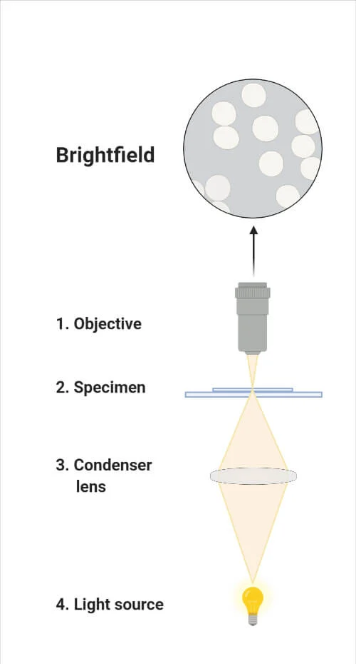
Brightfield Light Microscope
The confocal and super-resolution microscopes have revolutionized scientific research and diagnostics, but now let’s focus on the brightfield light microscope, which is a basic optical microscope commonly used in microbiology laboratories.
The brightfield light microscope produces a dark image against a bright background, allowing for the visualization of small organisms and cellular structures. Its components include the objective lens, eyepiece, condenser, and stage. Adequate lighting is crucial for obtaining high-resolution images with this microscope.
The brightfield light microscope works by using glass lenses to bend and magnify light, thereby enabling the observation of microscopic specimens. It’s particularly useful for studying stained samples, as the contrast between the specimen and the background is enhanced. However, it may not be suitable for transparent or unstained specimens.
Despite its simplicity, the brightfield light microscope remains an important tool in microbiology labs due to its versatility and affordability.
Phase Contrast Microscope
Now let’s delve into the topic of the phase contrast microscope.
This type of microscope is ideal for high contrast imaging and visualization of transparent specimens, such as microbial cultures and cell tissues.
High Contrast Imaging
High Contrast Imaging is achieved through the use of a Phase Contrast Microscope, which utilizes phase shifts to create high contrast images of unstained specimens. This type of microscope is ideal for viewing transparent specimens such as microbial cultures and cell tissues.
The components of a phase contrast microscope include a light source, collective lens, aperture, condenser, specimen, objective, phase plate, deflected light, and phase ring. The functioning of the phase contrast microscope involves converting light variations into an image.
The phase shifts caused by the specimen interact with the phase plate, creating contrast between different regions of the specimen. This allows for clear visualization of details that would otherwise be difficult to observe using traditional brightfield microscopy.
Transparent Specimen Visualization
To further explore the visualization of transparent specimens, we’ll now focus on the Phase Contrast Microscope, which utilizes phase shifts to create high contrast images of unstained specimens.
This microscope is ideal for viewing transparent specimens such as microbial cultures and cell tissues. The components of a phase contrast microscope include a light source, collective lens, aperture, condenser, specimen, objective, phase plate, deflected light, and phase ring.
The functioning of the phase contrast microscope involves converting light variations caused by the phase shifts of transparent specimens into an image. By manipulating the phase of the incident light, the microscope enhances the contrast between the specimen and its surroundings, allowing for detailed observation of unstained specimens.
The phase contrast microscope is a valuable tool in biological research and medical diagnostics, providing a non-destructive and non-invasive method for visualizing transparent specimens.
Dark-Field Microscope
The Dark-Field microscope is a specialized optical instrument used to visualize living unstained cells and the internal organs of larger eukaryotic cells. Unlike other types of microscopes, the Dark-Field microscope creates a dark background with a bright specimen, enhancing contrast and making it easier to observe transparent samples. This microscope is particularly useful for studying samples that are difficult to stain or those that may be damaged by the staining process.
The Dark-Field microscope operates on the principle of illuminating the specimen with a hollow cone of light. A dark-field stop is used to block the central portion of the light, allowing only the peripheral light rays to pass through the sample. These scattered rays are then captured by the objective lens, producing a bright image against a dark background.
The Dark-Field microscope consists of several components, including a light source, condenser, objective lens, and eyepiece. The light source is positioned at an angle to the condenser, which focuses the light into a cone shape. The objective lens collects the scattered light and forms an image, which is then magnified by the eyepiece for observation.
Fluorescent Microscope
Now let’s explore the fascinating world of fluorescent microscopy.
Fluorescent staining techniques allow for the visualization of specific molecules or structures within cells and tissues.
This technique has a wide range of applications in biomedical research, including studying cellular processes, identifying biomarkers, and investigating disease mechanisms.
Fluorescent Staining Techniques
Fluorescent staining techniques are widely utilized in conjunction with fluorescent microscopes for the visualization and analysis of specific molecules within biological samples. These techniques involve the use of fluorochromes, which are fluorescent dyes that bind to specific molecules of interest. By selectively staining these molecules, researchers can observe their location, distribution, and interactions within cells or tissues.
Fluorescent staining techniques offer several advantages over traditional staining methods. Firstly, they provide high specificity, allowing for the precise localization of targeted molecules. Secondly, they offer enhanced sensitivity, as the fluorescent signal can be amplified and detected with great precision. Additionally, fluorescent staining techniques enable multiplexing, where multiple molecules can be simultaneously visualized using different fluorochromes.
To perform fluorescent staining, the biological sample is incubated with the appropriate fluorochrome-conjugated antibody or probe. The fluorochrome specifically binds to the target molecule, resulting in fluorescence upon excitation with a specific wavelength of light. The sample is then visualized using a fluorescent microscope, which is equipped with filters to selectively capture the emitted fluorescent signal.
Applications in Biomedical Research
After discussing the advantages of fluorescent staining techniques in the visualization and analysis of specific molecules within biological samples, it’s important to explore the applications of fluorescent microscopes in the field of biomedical research.
Fluorescent microscopes play a critical role in various aspects of biomedical research, including the study of cellular processes, protein interactions, and disease mechanisms. These microscopes enable researchers to observe and monitor the localization and movement of specific molecules within living cells and tissues. By using fluorescent dyes or proteins that emit light of different colors, researchers can label specific structures or molecules of interest and visualize them under the microscope. This allows for the investigation of various biological processes, such as cell division, protein trafficking, and signal transduction.
Additionally, fluorescent microscopes are widely used in immunohistochemistry and immunofluorescence studies to detect and localize specific antigens or antibodies in tissue samples.
Applications of Dark-Field and Fluorescent Microscopes
Dark-field and fluorescent microscopes are invaluable tools in various fields of medical diagnostics and research, enabling the visualization and analysis of specific structures and processes within living cells and tissues.
Dark-field microscopy is particularly useful in studying living unstained cells and the internal organs of larger eukaryotic cells. By using a darkfield stop and a condenser lens, this type of microscope creates a hollow cone beam of light that illuminates the specimen from the sides. This technique produces a bright image against a dark background, making it easier to observe fine details and structures within the specimen.
On the other hand, fluorescent microscopy utilizes specific wavelengths of light to excite fluorochrome-stained specimens, causing them to emit fluorescence. This technique allows for the visualization of specific molecules or structures within the cell. For example, fluorescent tags can be used to label specific proteins or DNA sequences, enabling researchers to track their location and movement within the cell. This is particularly useful in studying cellular processes such as protein trafficking, cell division, and gene expression.
In medical diagnostics, dark-field and fluorescent microscopes are used to identify and analyze various diseases and conditions. Dark-field microscopy can be used to detect the presence of bacteria, such as Treponema pallidum, which causes syphilis.
Fluorescent microscopy, on the other hand, is commonly used in immunofluorescence assays to detect the presence of specific antibodies or antigens in patient samples. This technique is widely used in diagnosing infectious diseases, autoimmune disorders, and cancer.
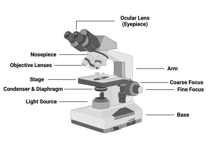
Parts of a Light Microscope
To continue our exploration of light microscopes, let’s now delve into the intricate components that make up this essential scientific tool.
A light microscope is composed of several parts that work together to enable the observation and magnification of small objects. The main components of a light microscope include the objective lens, eyepiece, condenser, and stage.
The objective lens is responsible for magnifying the specimen and capturing the image. It’s located at the bottom of the microscope and comes in various magnification powers.
The eyepiece, or ocular lens, is positioned at the top of the microscope and allows you to view the magnified image. The eyepiece typically has a magnification power of 10x.
The condenser is an important component that focuses light onto the specimen, ensuring optimal illumination. It’s located beneath the stage and can be adjusted to control the brightness and clarity of the image.
The stage provides a platform for placing the specimen and is equipped with mechanical controls that allow for precise movement and positioning.
Additional parts of a light microscope may include a diaphragm, which controls the amount of light passing through the condenser, and a coarse and fine focus adjustment knob, which allows for precise focusing of the specimen. Some microscopes also feature a built-in light source, such as an LED or halogen lamp, to provide illumination.
Understanding the different parts of a light microscope is crucial for proper usage and obtaining clear and accurate images. Each component plays a specific role in the functioning of the microscope, and their careful adjustment and alignment are essential for achieving optimal results.
Conclusion
In conclusion, light microscopes are essential tools for observing the microscopic world. By utilizing the principle of refraction and glass lenses, these microscopes greatly enhance our ability to detect and magnify small objects.
There are various types of light microscopes, each with its own unique functionality and specialized applications. From brightfield to phase contrast, dark-field, and fluorescent microscopes, these instruments offer a wide range of capabilities for medical diagnostics, research, and scientific exploration.
The parts of a light microscope work together to provide a comprehensive and detailed view of the wonders that lie beyond the naked eye.

Erzsebet Frey (Eli Frey) is an ecologist and online entrepreneur with a Master of Science in Ecology from the University of Belgrade. Originally from Serbia, she has lived in Sri Lanka since 2017. Eli has worked internationally in countries like Oman, Brazil, Germany, and Sri Lanka. In 2018, she expanded into SEO and blogging, completing courses from UC Davis and Edinburgh. Eli has founded multiple websites focused on biology, ecology, environmental science, sustainable and simple living, and outdoor activities. She enjoys creating nature and simple living videos on YouTube and participates in speleology, diving, and hiking.