
This post contains affiliate links. As an Amazon Associate I earn from qualifying purchases.
Monoclonal antibodies are produced in two main ways. The classic route is hybridoma technology: a mouse is immunized, its antibody making B cells are fused with immortal myeloma cells, and the one hybrid clone that makes the right antibody is grown in culture. The modern route is recombinant: the antibody genes are inserted into Chinese hamster ovary (CHO) cells, which secrete the antibody in steel or single use bioreactors before it is purified with Protein A chromatography.
That is the short answer. The long answer is one of the most elegant stories in biology, because every step solves a problem that once looked impossible. B cells make perfect antibodies but die in a dish. Cancer cells live forever but make nothing useful. Put the two together, and you get a cell that does both.
Key takeaways
- A monoclonal antibody is one identical antibody, copied from a single B cell clone, that binds one exact spot (epitope) on its target.
- Hybridoma technology was published by Georges Köhler and César Milstein in 1975 and earned them the 1984 Nobel Prize in Physiology or Medicine.
- The production path runs: immunize, fuse, select in HAT medium, screen, clone, scale up, purify.
- Most therapeutic antibodies today are recombinant and made in CHO cells. The rituximab label, for example, states it is produced in CHO suspension culture.
- The first therapeutic monoclonal antibody, muromonab CD3 (Orthoclone OKT3), was approved in 1986.
What is a monoclonal antibody?
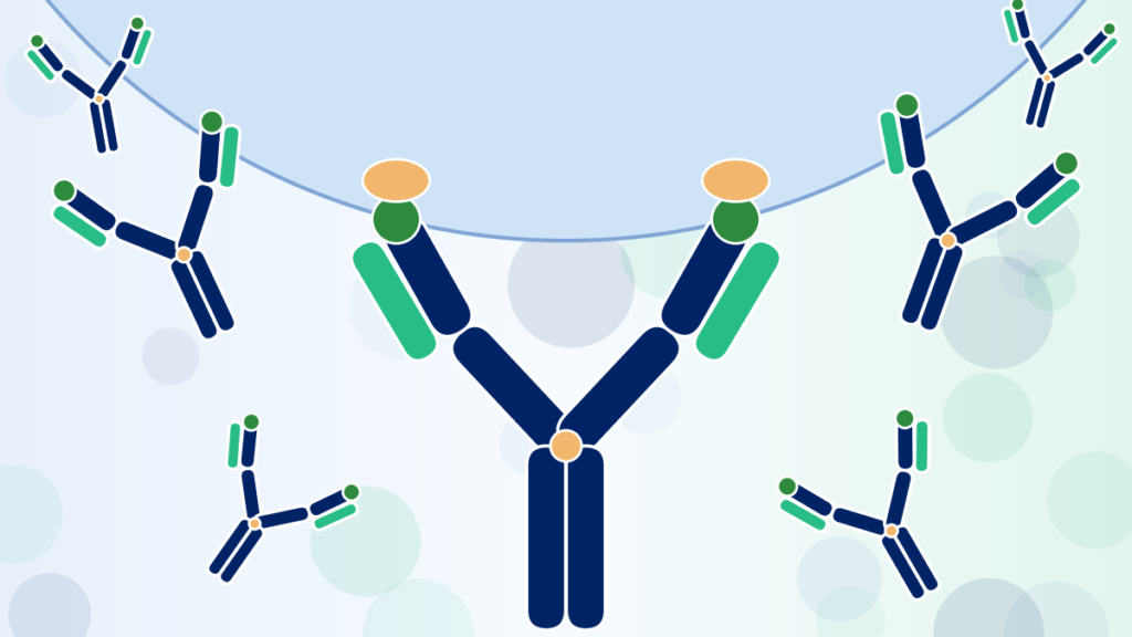
An antibody is a Y shaped protein made by B cells. The two tips of the Y carry variable regions that grip a specific molecular shape, the epitope. The stem, called the Fc region, tells the rest of the immune system what to do once the antibody has latched on.
When you get an infection, hundreds of different B cells respond at once, each making a slightly different antibody. That mixture is polyclonal. A monoclonal antibody is the opposite: every molecule in the vial is a copy of one antibody from one B cell lineage. That sameness is the whole point. It makes the antibody predictable enough to use as a lab reagent, a diagnostic test or a drug.
How monoclonal antibodies are produced with hybridoma technology
Hybridoma technology is still the method most biology students learn first, and it is still used to discover new antibodies. Here is how it works, step by step.
Step 1: Immunize the animal
A mouse is injected with the target, called the antigen. It can be a purified protein, a peptide or whole cells. Over several weeks of booster shots, the mouse’s immune system selects and multiplies the B cells whose antibodies bind that antigen best. Researchers test small blood samples to confirm the immune response is strong before moving on.
Step 2: Harvest B cells from the spleen
The spleen is packed with B cells, including the ones trained against the antigen. It is removed and broken up into a suspension of single cells. These B cells are the antibody factories, but they have a fatal flaw: outside the body they stop dividing and die within days.
Step 3: Fuse them with myeloma cells
Myeloma cells are cancerous plasma cells that divide forever. The lines used for fusion have been chosen because they make no antibody of their own. Mixing spleen cells and myeloma cells with polyethylene glycol (PEG) makes their membranes merge. A small fraction of the pairs become hybridomas: single cells that carry the B cell’s antibody genes and the myeloma cell’s immortality.
Step 4: Select hybridomas in HAT medium
After fusion the dish holds a messy crowd: unfused B cells, unfused myeloma cells and the rare hybrids. The cleanup is done by the growth medium itself. HAT medium contains hypoxanthine, aminopterin and thymidine. Aminopterin blocks the main pathway cells use to build DNA, so they must use a backup pathway that needs the enzyme HGPRT. The myeloma lines lack HGPRT and die. Unfused B cells die on their own. Only hybridomas, which inherit HGPRT from the B cell and immortality from the myeloma, survive. If you want to see how a growth medium is built to favor some cells over others, my guide to microbial growth media preparation walks through the same logic in microbiology.
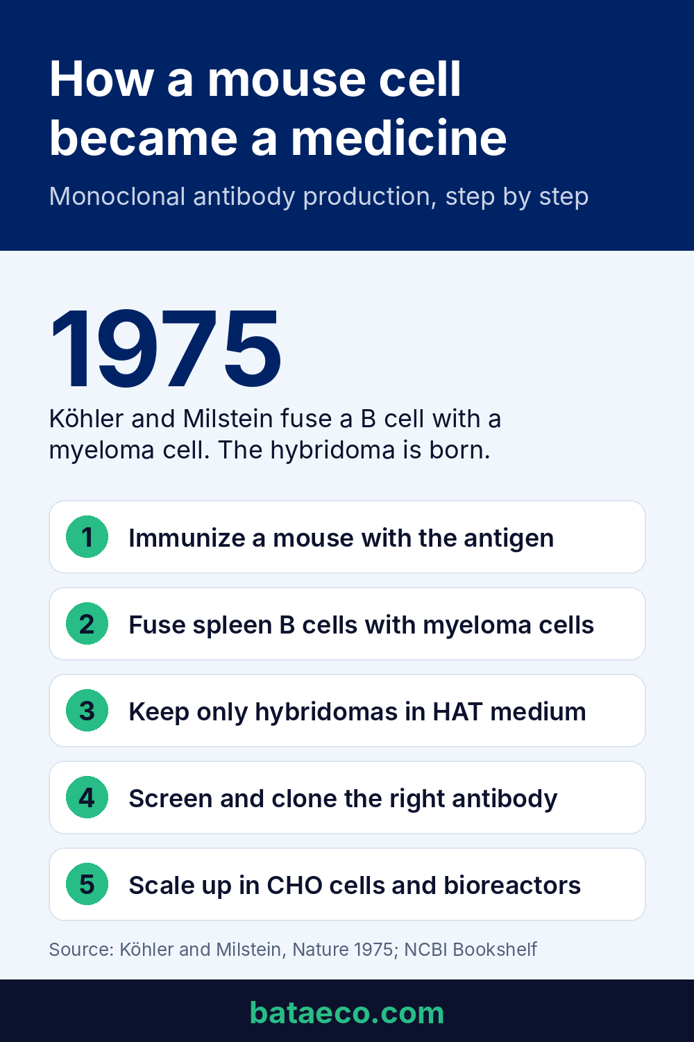
Step 5: Screen for the right antibody
Surviving hybridomas are spread across 96 well plates, and the liquid above each well is tested, most often by ELISA, to see whether it contains antibody that binds the antigen. Many wells are blank or bind the wrong thing. The few positive wells are the candidates.
Step 6: Clone by limiting dilution
A positive well can still hold several different hybridomas. To make the antibody truly monoclonal, the cells are diluted so far that, on average, a well receives one cell or none. Each colony that grows from a single cell is a clone, and every antibody it secretes is identical.
This is the step where counting matters. During a three month lab internship, I spent long hours counting cells under the microscope, and it taught me how much quiet precision hides behind a word like “dilute”. Get the cell count wrong and a well that should hold one cell holds three, and your antibody is no longer monoclonal.
Step 7: Scale up and harvest
The winning clone is expanded in flasks and then in larger culture systems, and the antibody is collected from the culture liquid. An older shortcut grew hybridomas inside the abdomen of mice, the ascites method. A National Research Council committee advised that in vitro methods should be the first choice, with ascites used only when culture is not reasonable, and today most production happens in culture.
Why mouse antibodies had to be made human
Here is the honest twist in the story. Hybridomas solved production, but the antibodies they made were mouse proteins. Injected into people, they could trigger the patient’s immune system to make antibodies against the drug itself, which shortened how long it worked and caused reactions.
The fix was genetic engineering. Scientists cut the antigen binding regions out of the mouse antibody and grafted them onto a human antibody frame. Under the older naming system you can still read this history in drug names:
- Murine (mouse): names ending in “omab”, such as ibritumomab.
- Chimeric: mouse variable regions on a human constant region, names ending in “ximab”, such as rituximab.
- Humanized: only the smallest binding loops are mouse, names ending in “zumab”, such as trastuzumab.
- Fully human: no mouse sequence, names ending in “umab”, such as adalimumab.
Fully human antibodies came from two directions: mice engineered to carry human antibody genes, and phage display, a method that shows huge libraries of human antibody fragments on the surface of viruses so the best binders can be fished out. George Smith and Gregory Winter shared the 2018 Nobel Prize in Chemistry for phage display work, and adalimumab, one of the best known antibody drugs, was discovered this way.
Case study: what 156 approved antibody drugs reveal
I wanted to know whether the move from mouse to human antibodies is a real pattern or just a neat textbook story. So I went through every antibody therapeutic in The Antibody Society’s list of US and EU approvals and sorted each one by origin (mouse, chimeric, humanized or fully human, as the list records its format) and by the decade of its first approval.
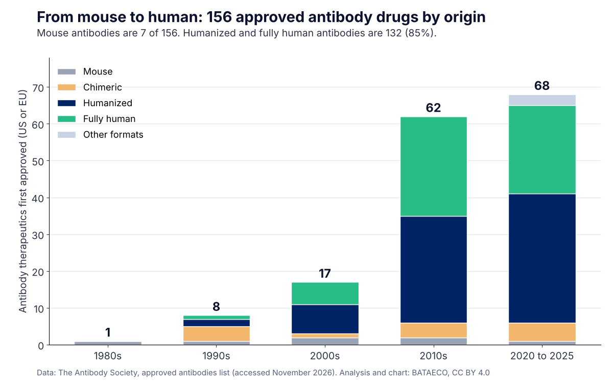
- 156 antibody therapeutics had a first US or EU approval in the list, starting with muromonab CD3 in 1986.
- Only 7 are mouse antibodies, about 4 percent. Humanized antibodies lead with 74 (47 percent), followed by fully human with 58 (37 percent), chimeric with 14 (9 percent) and 3 newer bispecific formats.
- Since 2000, just 5 of 147 approvals were mouse antibodies. Humanization and fully human discovery replaced them almost completely.
- The 2020s are already ahead of the 2010s: 68 first approvals from 2020 to 2025, compared with 62 in the whole previous decade.
What this means for production is quiet but enormous. Almost every modern antibody drug is humanized or human, which means it is manufactured from a known gene sequence in a recombinant cell line. The hybridoma did not disappear, it moved upstream into discovery, and the real production work moved into CHO cells and bioreactors.
Method: for each product I used the earliest of its first EU or first US approval year and the format listed for it. Products still under review were excluded.
How therapeutic monoclonal antibodies are produced today
Once you know the antibody’s gene sequence, you no longer need the hybridoma. Modern antibody drugs are recombinant: the genes for the heavy and light chains are written into a DNA vector and delivered into a production cell line. It is the same basic idea as bacterial transformation, where a plasmid carries new instructions into a cell, only scaled up to mammalian cells and to a level of control that drug regulators demand.
Cell line development in CHO cells
The workhorse is the Chinese hamster ovary cell. CHO cells grow in suspension, tolerate industrial conditions, and attach sugar chains to proteins in a way close enough to human cells that the antibody behaves properly in the body. The prescribing information for the rituximab biosimilar Ruxience, for example, states it is produced in Chinese hamster ovary suspension culture. After transfection, thousands of cell clones are screened to find one stable, high producing line, and that single clone becomes the master cell bank for the product’s entire life.
Upstream: growing cells in bioreactors
Cells from the bank are expanded step by step into production bioreactors, from stainless steel tanks of thousands of liters to single use bag reactors. Most processes run as fed batch: nutrients are added during a run of around two weeks while temperature, pH, dissolved oxygen and cell density are tracked continuously. The cells secrete the antibody into the culture liquid, so the product never has to be extracted from inside them.
Downstream: purification
Purification turns cloudy culture broth into an injectable medicine. A typical sequence looks like this:

- Harvest and clarification: centrifugation and depth filtration remove cells and debris.
- Protein A capture: a column coated with Protein A, a bacterial protein that grips the Fc region of antibodies, pulls the antibody out of the broth in one step.
- Low pH viral inactivation: a short hold at acidic pH inactivates enveloped viruses.
- Polishing chromatography: ion exchange steps remove host cell proteins, DNA and clumped antibody.
- Viral filtration: nanofilters remove any remaining virus particles.
- Formulation: ultrafiltration exchanges the antibody into its final buffer at the right concentration before sterile filling.
Hybridoma vs recombinant production at a glance
| Hybridoma | Recombinant (CHO) | |
|---|---|---|
| Source of antibody genes | Fused mouse B cell | Known DNA sequence in a vector |
| Typical antibody type | Mouse | Chimeric, humanized or fully human |
| Main use today | Discovery and research reagents | Therapeutic antibody drugs |
| Sequence known and fixed | Not always | Yes, from the start |
| Risk of drift between batches | Higher, hybridomas can lose expression | Lower, controlled cell banks |
How long does it take to make monoclonal antibodies?
For a research antibody, immunization through cloning usually takes a few months, with several more weeks to produce and test useful amounts. A therapeutic antibody is a different scale entirely. Discovery, engineering, cell line development, safety studies and clinical trials together commonly stretch to around a decade before approval.
From one Nobel idea to a whole class of medicines
The first therapeutic monoclonal antibody, muromonab CD3, was approved in 1986 to prevent kidney transplant rejection. The Antibody Society lists 156 antibody therapeutics with a US or EU approval in its approved antibodies list. Every one of them traces back to a 1975 experiment in which two scientists asked a simple question: what if we could make a cell that never forgets the antibody it was trained to make?
Books and tools for learning antibody production
If you are studying immunology or starting work in an antibody lab, these are the resources I would reach for.
- Antibodies: A Laboratory Manual (Cold Spring Harbor): the bench reference for immunization, hybridoma work and antibody purification. Best for students and technicians who need actual protocols.
- Janeway’s Immunobiology: the clearest explanation of how B cells and antibodies work in the body. Best for undergraduates who want the biology behind the methods.
- Kuby Immunology: a popular course textbook with strong figures on antibody structure and hybridoma technology. Best as an exam companion.
- A hemocytometer: the glass counting chamber used to measure cell density before dilution and cloning. Best for anyone practicing cell culture skills.
Frequently asked questions
What are the steps of monoclonal antibody production?
With hybridoma technology the steps are: immunize a mouse, harvest spleen B cells, fuse them with myeloma cells, select hybridomas in HAT medium, screen by ELISA, clone by limiting dilution, then scale up and purify. Recombinant production replaces the first steps with cloning the antibody genes into CHO cells.
What is the downside of monoclonal antibodies?
They are expensive to make, they usually must be given by injection or infusion because the stomach digests proteins, they need cold storage, and the body can sometimes form antibodies against them, especially older mouse antibodies.
How are primary antibodies made for research?
Primary antibodies used in Western blots, ELISA and staining are made the same two ways: monoclonal ones from hybridomas or recombinant cell lines, and polyclonal ones by immunizing rabbits, goats or other animals and collecting antibodies from their blood.
Which cells are used to produce therapeutic monoclonal antibodies?
Most therapeutic antibodies are produced in Chinese hamster ovary (CHO) cells. Other mammalian lines such as NS0 and SP2/0 mouse myeloma cells have also been used.
This article explains how antibodies are made for education. It is not medical advice about any antibody treatment.

Erzsebet (Eli) Frey is a biologist with a BSc in Biology and an MSc in Ecology from the University of Belgrade. She has field and lab experience across Asia, the Middle East, Europe and South America and uses Python and machine learning on biological data. Based in Serbia, she writes BATAECO and also runs Simply Ecologist and Eco&Stylish.