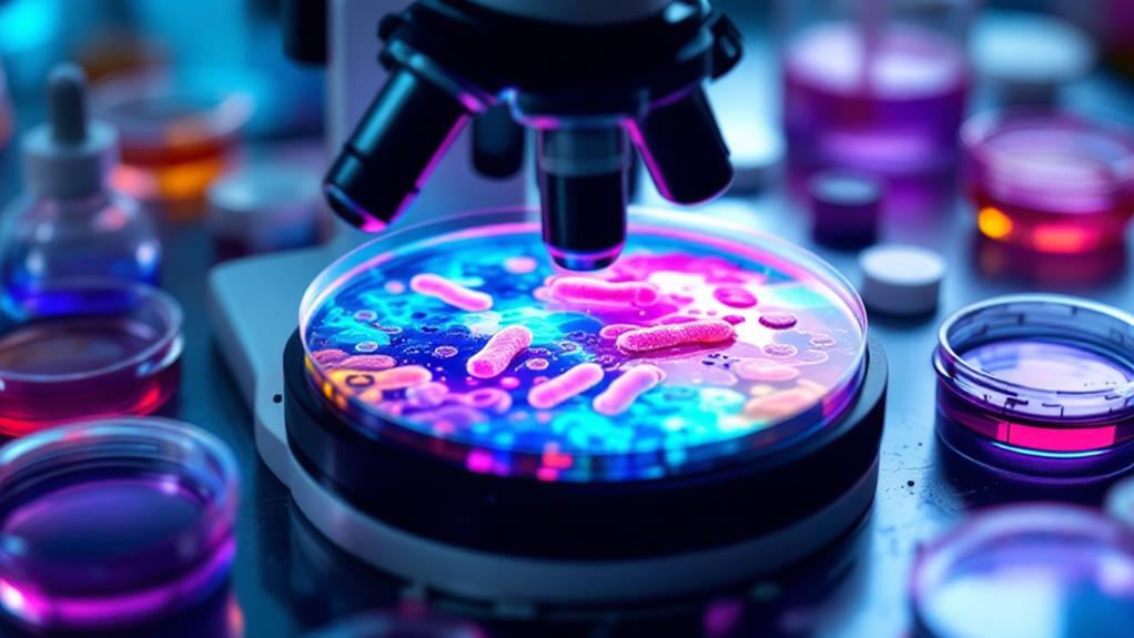
Microbiology staining techniques are crucial tools for identifying and studying microorganisms. You'll encounter various methods, each serving a specific purpose. Gram staining differentiates bacteria based on cell wall composition, while acid-fast staining discloses waxy cell walls. Simple and negative staining offer quick ways to visualize microbial shape and size. For more detailed observations, special techniques like flagella and endospore staining come into play. Advanced methods include fluorescent staining for specific cellular components and critical staining for observing living organisms. These techniques form the backbone of microbial identification and research, opening up a fascinating world of microscopic life. Exploring each method will disclose the intricacies of bacterial structures and behaviors.
Gram Staining
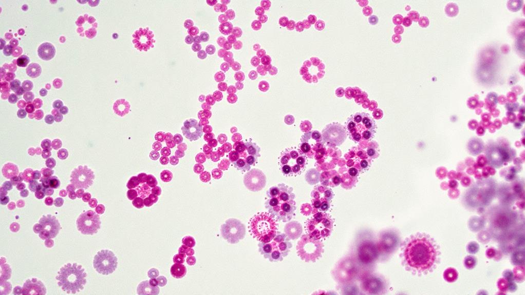
Gram staining, a cornerstone of bacterial identification, is a differential staining technique that distinguishes between gram-positive and gram-negative bacteria. It's named after Hans Christian Gram, who developed the method in 1884. The process relies on the structural differences in bacterial cell walls to create a visual contrast between the two types.
To perform a Gram stain, you'll need to follow a specific sequence of steps. First, you'll prepare a thin smear of bacteria on a glass slide and heat-fix it. Then, you'll apply crystal violet, the primary stain, which colors all bacteria purple. Next, you'll add Gram's iodine, a mordant that forms a complex with the crystal violet.
The critical step comes next: decolorization with alcohol or acetone. Gram-positive bacteria retain the crystal violet-iodine complex, staying purple. Gram-negative bacteria, however, lose this complex and become colorless. Finally, you'll apply a counterstain, usually safranin, which turns gram-negative bacteria pink or red.
The key to interpreting Gram stains lies in understanding cell wall composition. Gram-positive bacteria have a thick peptidoglycan layer that traps the crystal violet-iodine complex. Gram-negative bacteria have a thin peptidoglycan layer and an outer membrane, allowing the complex to wash away during decolorization.
You'll find Gram staining invaluable in clinical microbiology for rapid bacterial identification and guiding initial antibiotic therapy. It's also useful in research settings for characterizing bacterial isolates. Remember, while Gram staining is a powerful tool, it's not infallible. Some bacteria are Gram-variable or don't stain well, requiring additional tests for accurate identification.
Acid-Fast Staining
Acid-fast staining stands out as another vital technique in the microbiologist's toolkit. This method is significant for identifying bacteria with cell walls rich in mycolic acids, particularly members of the Mycobacterium genus, including M. tuberculosis and M. leprae.
You'll find that acid-fast bacteria resist decolorization by acid-alcohol solutions after being stained with primary dyes. This unique characteristic is due to their waxy cell walls, which contain high amounts of lipids. The process involves heating the smear with carbol fuchsin, a pink-red dye that penetrates the cell wall. When you apply an acid-alcohol solution, it won't remove the dye from acid-fast bacteria, while non-acid-fast bacteria will decolorize.
To perform acid-fast staining, you'll need to follow these steps:
- Prepare and heat-fix the bacterial smear
- Flood the slide with carbol fuchsin and heat it
- Rinse with water
- Decolorize with acid-alcohol
- Counterstain with methylene blue
After staining, you'll observe acid-fast bacteria as bright red rods against a blue background. Non-acid-fast bacteria and other cellular material will appear blue.
It's important to note that there are variations of the acid-fast stain, such as the Ziehl-Neelsen and Kinyoun methods. These techniques differ slightly in their procedures but achieve similar results. You might also encounter fluorescent acid-fast stains, which use fluorescent dyes instead of carbol fuchsin, offering increased sensitivity and easier visualization under a fluorescence microscope.
Negative Staining
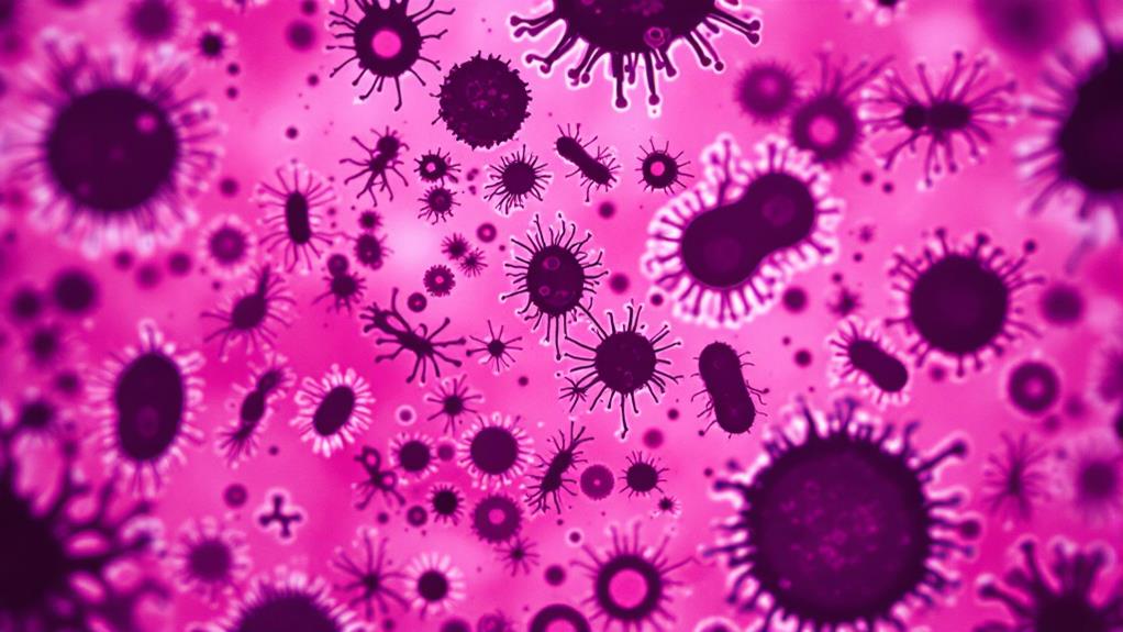
Unlike other staining techniques, negative staining stands out for its simplicity and unique approach. You'll find it's particularly useful for visualizing the overall shape and structure of microorganisms, especially those that are difficult to stain using conventional methods. In negative staining, you're not actually staining the specimen itself, but rather the background around it.
To perform a negative stain, you'll start by placing a drop of the microbial suspension on a microscope slide. Next, you'll add a drop of dark staining solution, such as nigrosin or India ink, to the suspension. After mixing the two drops, you'll spread the mixture thinly across the slide using the edge of another slide. Once it's dry, you're ready to examine the specimen under a microscope.
When you look through the microscope, you'll see light-colored microorganisms against a dark background. This contrast allows you to observe the cell's outer shape and any external structures like flagella or capsules. It's particularly useful for examining bacteria that are too small to see clearly with other staining methods.
You'll find negative staining especially valuable when working with live specimens, as it doesn't kill the microorganisms. This means you can observe their natural movements and behavior. It's also a quick technique that doesn't require heat fixation or multiple staining steps.
However, you should be aware that negative staining won't provide information about internal cellular structures. For that, you'll need to use other staining techniques. Despite this limitation, negative staining remains a valuable tool in your microbiology toolkit, offering a straightforward way to visualize microbial morphology.
Simple Staining
With simple staining, you'll encounter one of the most basic yet effective techniques in microbiology. This method uses a single dye to color bacterial cells, making them visible under a microscope. It's a quick and easy way to observe the general shape, size, and arrangement of microorganisms.
To perform a simple stain, you'll need a clean glass slide, a bacterial sample, and a basic dye. Common dyes include methylene blue, crystal violet, or safranin. First, prepare a thin smear of your bacterial sample on the slide and let it air dry. Then, heat-fix the smear by passing it through a flame a few times. This step helps the cells adhere to the slide.
Next, you'll apply the chosen dye to the fixed smear and let it sit for about a minute. Afterward, gently rinse the slide with water to remove excess dye. Blot the slide dry with bibulous paper, and it's ready for microscopic examination.
When you look at your simple-stained sample under a microscope, you'll see that all the bacterial cells appear the same color. This uniformity allows you to focus on the morphology and arrangement of the cells. You can easily distinguish between cocci (spherical), bacilli (rod-shaped), or spirilla (spiral-shaped) bacteria.
Simple staining has its limitations, though. It won't provide information about cell wall composition or internal structures. For more detailed observations, you'll need to use differential staining techniques. However, simple staining remains an essential tool in microbiology labs for quick bacterial identification and morphology studies.
Flagella Staining
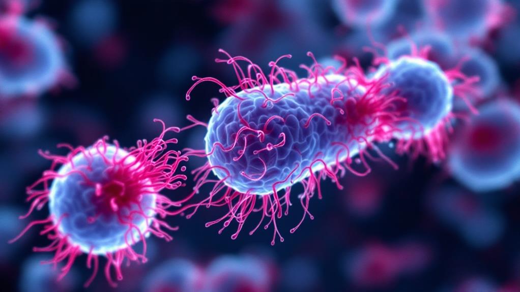
Flagella staining presents a unique challenge in microbiology due to the delicate nature of these bacterial appendages. Unlike other cellular structures, flagella are extremely thin and fragile, making them difficult to visualize under a standard microscope. To overcome this obstacle, you'll need to employ specialized staining techniques that increase the thickness of the flagella, allowing them to become visible.
The most common method for flagella staining is the Leifson technique. In this procedure, you'll first prepare a suspension of the bacterial culture on a clean glass slide. Next, you'll apply a mordant solution containing tannic acid, which helps the stain adhere to the flagella. After allowing the mordant to act for a few minutes, you'll add the primary stain, typically silver nitrate. The silver ions will precipitate along the flagella, effectively increasing their diameter.
It's vital to handle the slide gently throughout the process, as any rough treatment can break or detach the flagella. Once you've completed the staining, you'll rinse the slide carefully and allow it to air dry. Under the microscope, you'll be able to observe the flagella as dark, wavy structures extending from the bacterial cells.
Remember that flagella staining requires patience and practice. You may need to adjust the staining time or concentration of reagents to achieve ideal results. It's also important to note that not all bacteria possess flagella, so you should choose appropriate species for this technique. By mastering flagella staining, you'll gain valuable insights into bacterial motility and morphology.
Endospore Staining
Endospore staining frequently poses a challenge for microbiologists due to the resilient nature of bacterial endospores. These dormant structures are highly resistant to conventional staining methods, making them difficult to visualize under a microscope. To overcome this obstacle, you'll need to use specialized endospore staining techniques.
The most common method for endospore staining is the Schaeffer-Fulton technique. You'll start by preparing a bacterial smear on a microscope slide and heat-fixing it. Next, you'll flood the slide with malachite green stain and apply heat for several minutes. This process forces the dye into the tough endospore coat. After rinsing, you'll counterstain with safranin, which colors the vegetative cells red.
When you examine the slide under a microscope, you'll see red vegetative cells with green endospores inside them. The contrast between the two colors makes it easy to identify and locate the endospores within the bacterial cells.
Another technique you might use is the Wirtz-Conklin method. This approach uses carbol fuchsin as the primary stain and methylene blue as the counterstain. The process is similar to the Schaeffer-Fulton technique, but it results in red endospores against blue vegetative cells.
It's essential to note that not all bacteria form endospores. Species like Bacillus and Clostridium are known endospore formers, while many other bacterial genera don't produce these structures. By mastering endospore staining techniques, you'll be able to identify and study these important bacterial structures effectively.
Fluorescent Staining
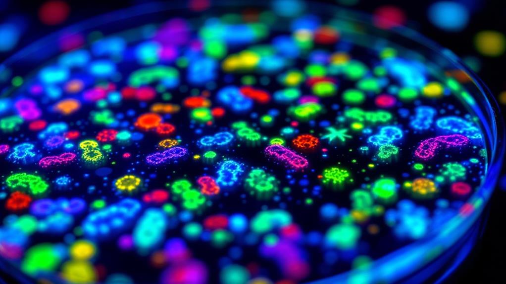
Illuminating the microscopic world, fluorescent staining techniques have revolutionized microbiology research. These methods allow you to visualize specific cellular components, proteins, or nucleic acids with remarkable clarity. By using fluorescent dyes or fluorophore-labeled antibodies, you can observe targeted structures that emit light when excited by specific wavelengths.
To perform fluorescent staining, you'll first need to fix and permeabilize your sample. This step guarantees that the fluorescent molecules can access their targets within the cell. You'll then apply the fluorescent stain or antibody, allowing it to bind to the structures of interest. After washing away excess stain, you can observe your sample under a fluorescence microscope.
One popular technique is FISH (Fluorescence In Situ Hybridization), which uses fluorescent probes to detect specific DNA or RNA sequences. This method's particularly useful for identifying and localizing genetic material within cells or tissues.
Another common approach is immunofluorescence, where you'll use fluorescently labeled antibodies to detect specific proteins. This technique's invaluable for studying protein localization and interactions within cells.
Fluorescent staining isn't limited to fixed samples. You can also use live-cell imaging techniques with fluorescent dyes that don't harm living cells. This allows you to observe dynamic processes in real-time.
When working with multiple fluorescent stains, you'll need to carefully select dyes with non-overlapping emission spectra. This enables you to simultaneously visualize different cellular components in a single sample, providing a wealth of information about cellular structure and function.
Vital Staining
While fluorescent staining often requires fixed samples, essential staining allows you to observe living microorganisms in real-time. This technique is invaluable when you need to study cellular processes, assess viability, or examine the behavior of live cells without killing them.
You'll find that essential stains are typically non-toxic and don't interfere with cellular functions. Common essential stains include methylene blue, which you can use to observe yeast cells, and trypan blue, which helps you distinguish between live and dead cells. When you're working with trypan blue, live cells will exclude the dye, appearing clear, while dead cells absorb it and turn blue.
To perform essential staining, you'll need to prepare a fresh sample of your microorganisms. Mix a small amount of the stain with your sample on a microscope slide. It's important to work quickly, as some essential stains can become toxic to cells over time. You'll then observe the sample under a microscope, where you can see various cellular structures and processes in action.
One of the most exciting applications of essential staining is in studying bacterial motility. You can use a technique called hanging drop preparation, where you suspend a drop of stained bacterial culture from a coverslip. This allows you to observe the movement of live bacteria in three dimensions.
Remember that essential staining has its limitations. Some cellular structures may not be visible, and the stain's effectiveness can vary depending on the organism. However, it remains an indispensable tool in your microbiology toolkit, offering unique insights into living microorganisms that other staining methods can't provide.

Erzsebet Frey (Eli Frey) is an ecologist and online entrepreneur with a Master of Science in Ecology from the University of Belgrade. Originally from Serbia, she has lived in Sri Lanka since 2017. Eli has worked internationally in countries like Oman, Brazil, Germany, and Sri Lanka. In 2018, she expanded into SEO and blogging, completing courses from UC Davis and Edinburgh. Eli has founded multiple websites focused on biology, ecology, environmental science, sustainable and simple living, and outdoor activities. She enjoys creating nature and simple living videos on YouTube and participates in speleology, diving, and hiking.

