
You'll be using a microscope to examine microorganisms, and accurate technique is essential to obtain reliable results. First, set up your microscope by calibrating the stage and selecting the right objective lens for your sample. Handle your samples carefully to prevent contamination, and process them for microscopic examination using proper staining and fixing techniques. Next, adjust the microscope's focus to capture microorganisms' intricate details. Use the fine adjustment knob to refine the focus, and pay attention to microorganisms' movement and behavior. As you master these techniques, you'll uncover the intricacies of microbiology, and discover the secrets hidden in plain sight. Now, take a closer look.
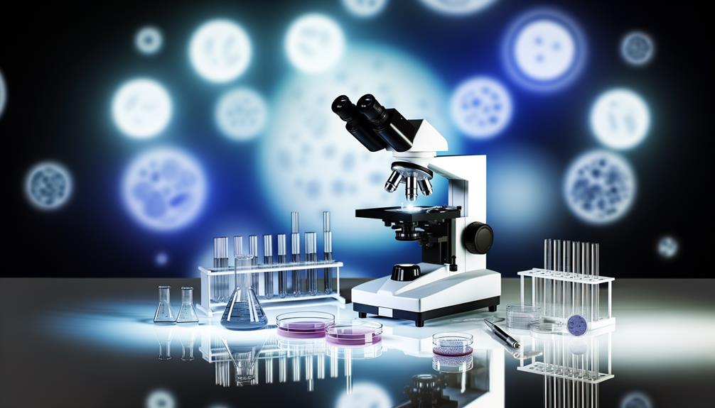
Understanding Microscope Components
You'll be working with several key components when using a microscope, including the base, stage, and optical system.
The base provides stability and support for the instrument, while the stage is where you'll place your specimen slides.
The optical system, comprising the objective lenses, ocular lenses, and light source, is responsible for magnifying and illuminating your sample.
The objective lenses, typically ranging from 4x to 100x magnification, are located just above the stage. You'll choose the appropriate objective lens based on the level of magnification required for your sample.
The ocular lenses, usually 10x or 15x, are found in the eyepieces and further magnify the image.
The light source, often a built-in LED or halogen lamp, transmits light through the specimen, allowing you to visualize it.
Understanding the roles of each component is vital for proper microscope operation.
Familiarize yourself with the location and function of each part to confirm you're using the microscope efficiently and effectively.
Preparing Microbiology Samples
To prepare microbiology samples for microscopic examination, carefully collect and process specimens to prevent contamination and preserve the integrity of the sample.
You'll need to follow proper protocols for sample collection, handling, and storage to maintain the quality of your results. When collecting samples, use sterile equipment and techniques to minimize the risk of contamination.
If you're working with clinical samples, be sure to follow proper biosafety protocols to protect yourself and others.
Once you've collected your sample, you'll need to process it for microscopic examination. This may involve staining the sample to enhance visibility, fixing the sample to preserve its morphology, or preparing a smear or section for observation.
To guarantee accurate and reliable results, follow established protocols for sample preparation. Remember to handle your samples gently to prevent damage and to store them properly to maintain their integrity.
Selecting the Right Objective Lens
With your microbiology sample prepared, it's now time to focus on the microscope's objective lenses, which play a critical role in resolving the fine details of your specimen.
You'll need to select the right objective lens to achieve maximum magnification and resolution. Typically, microscopes have multiple objective lenses, each with a specific magnification power, such as 4x, 10x, 40x, and 100x.
When choosing an objective lens, consider the type of microbiology sample you're working with and the level of detail you need to observe.
For example, a 4x or 10x objective lens is suitable for observing larger structures, like bacterial colonies or fungal hyphae. For higher magnification, you'll need a 40x or 100x objective lens, which is ideal for observing smaller structures, like individual bacterial cells or viral particles.
Remember to always start with a lower magnification lens and gradually move to higher magnification lenses to verify you're focusing on the correct area of the specimen.
Calibrating the Microscope Stage
The microscope stage, which holds your specimen, requires precise calibration to guarantee accurate and consistent results.
You'll need to adjust the stage's X-Y axis to confirm it's properly aligned with the objective lenses. Start by rotating the stage's adjustment knobs counterclockwise to loosen the stage's holding mechanism. Next, gently move the stage to the desired position, making sure it's centered and even. You may need to adjust the stage's height using the coarse adjustment knob to achieve perfect focus.
Once you've positioned the stage, tighten the holding mechanism by rotating the adjustment knobs clockwise. Verify that the stage is securely locked in place by gently tugging on it.
Now, using the fine adjustment knob, make any necessary fine-tuned adjustments to the stage's position. Remember to work slowly and deliberately, as even slight misalignments can compromise your results.
With the stage properly calibrated, you'll be able to achieve precise, high-magnification observations of your microorganisms.
Focusing on Microorganisms
Adjusting the microscope's focus to capture microorganisms' intricate details requires a combination of precise stage calibration and deliberate manipulation of the focus knobs.
You'll need to rotate the coarse adjustment knob to bring the microorganisms into rough focus. Then, use the fine adjustment knob to refine the focus, making subtle adjustments until the image is sharp. Be cautious not to over- or under-focus, as this can lead to distorted or blurry images.
As you focus, pay attention to the microorganisms' movement and behavior. You may need to adjust the microscope's illumination to optimize contrast and visibility.
For instance, you can adjust the condenser aperture to reduce glare or switch to a different objective lens to enhance resolution. Remember to work methodically, making small adjustments and checking the image regularly to avoid losing your focus.
Identifying Microbial Morphology
You'll distinguish microbial morphology by examining the shape, size, and arrangement of cells, which can reveal essential information about their functionality and behavior.
This examination is vital in microbiology, as it helps you understand how microorganisms interact with their environment and respond to different stimuli.
When observing microbial morphology, pay attention to the cell shape, which can be spherical, rod-shaped, or spiral.
Note the size of the cells, as it can indicate their metabolic activity.
The arrangement of cells is also important, as it can suggest whether the microorganism is a single-celled or multicellular organism.
You may observe cells that are clustered together, forming colonies, or those that are scattered individually.
Additionally, observe the presence of appendages, such as flagella, which can indicate motility.
Using Staining Techniques Effectively
By applying appropriate staining techniques, you can selectively highlight specific structures or features within microbial cells, enabling you to gather more detailed information about their morphology and behavior.
This allows you to differentiate between various microorganisms, identify specific characteristics, and understand their interactions with their environment.
When using staining techniques, selecting the right stain for the desired outcome is vital.
For instance, Gram staining is commonly used to distinguish between Gram-positive and Gram-negative bacteria, while acid-fast staining is used to detect Mycobacterium tuberculosis.
You should also consider the type of microscopy you're using, as some stains are optimized for brightfield microscopy, while others are better suited for fluorescence microscopy.
To verify reproducibility and accuracy, adhering to standardized staining protocols is key.
This includes using the correct concentrations, incubation times, and washing procedures to prevent artifacts and non-specific binding.
Capturing and Analyzing Images
With your stained samples prepared, you're now ready to capture high-quality images using your microscope, which will enable you to analyze the morphology and behavior of microbial cells in greater detail.
To do this, you'll need to adjust the microscope's settings to optimize image quality. Start by adjusting the lighting to minimize glare and promote even illumination. Next, focus the microscope using the coarse and fine focus knobs to achieve a sharp image.
Once you've achieved a clear image, use the camera or smartphone adapter to capture the image. You can use image analysis software to enhance and measure the images, allowing you to quantify features such as cell size, shape, and distribution.
When analyzing the images, take note of the morphology of the microbial cells, including their shape, size, and arrangement. You may also observe features such as flagella, capsules, or spores.
Avoiding Common Microscopy Errors
When examining microbial cells under the microscope, be aware of common errors that can compromise the accuracy of your observations and analyses, as it's vital to verify the reliability of your results.
One common mistake is incorrect microscope setup, which can lead to poor image quality and inaccurate measurements. Make sure to follow the manufacturer's instructions and calibrate the microscope properly before use.
Another error to avoid is inadequate sample preparation, which can result in misleading observations. Confirm that your samples are properly stained, fixed, and mounted to prevent distortion or degradation of the sample. Additionally, be cautious of contamination and handling errors, which can introduce artifacts or alter the sample's composition.
You should also be mindful of your own biases and assumptions when interpreting your observations. Avoid making premature conclusions or over-interpreting your data, as this can lead to incorrect conclusions.
Maintaining a Sterile Microscope Environment
To prevent contamination and maintain the integrity of your samples, you must maintain a sterile microscope environment by controlling temperature, humidity, and air quality, and by implementing proper cleaning and disinfection protocols.
This is vital in microbiology, where even the slightest contamination can compromise the accuracy of your results.
You should verify that your microscope is located in a room with minimal air traffic and away from direct sunlight, which can cause temperature fluctuations.
The ideal temperature range for a microscope environment is between 20°C to 25°C, with a relative humidity of 40% to 60%.
Regularly cleaning and disinfecting the microscope, as well as the surrounding surfaces, is essential.
You can use 70% ethanol or isopropanol for disinfection.
Additionally, you should wear gloves and a lab coat to prevent introducing external contaminants into the environment.

Erzsebet Frey (Eli Frey) is an ecologist and online entrepreneur with a Master of Science in Ecology from the University of Belgrade. Originally from Serbia, she has lived in Sri Lanka since 2017. Eli has worked internationally in countries like Oman, Brazil, Germany, and Sri Lanka. In 2018, she expanded into SEO and blogging, completing courses from UC Davis and Edinburgh. Eli has founded multiple websites focused on biology, ecology, environmental science, sustainable and simple living, and outdoor activities. She enjoys creating nature and simple living videos on YouTube and participates in speleology, diving, and hiking.