
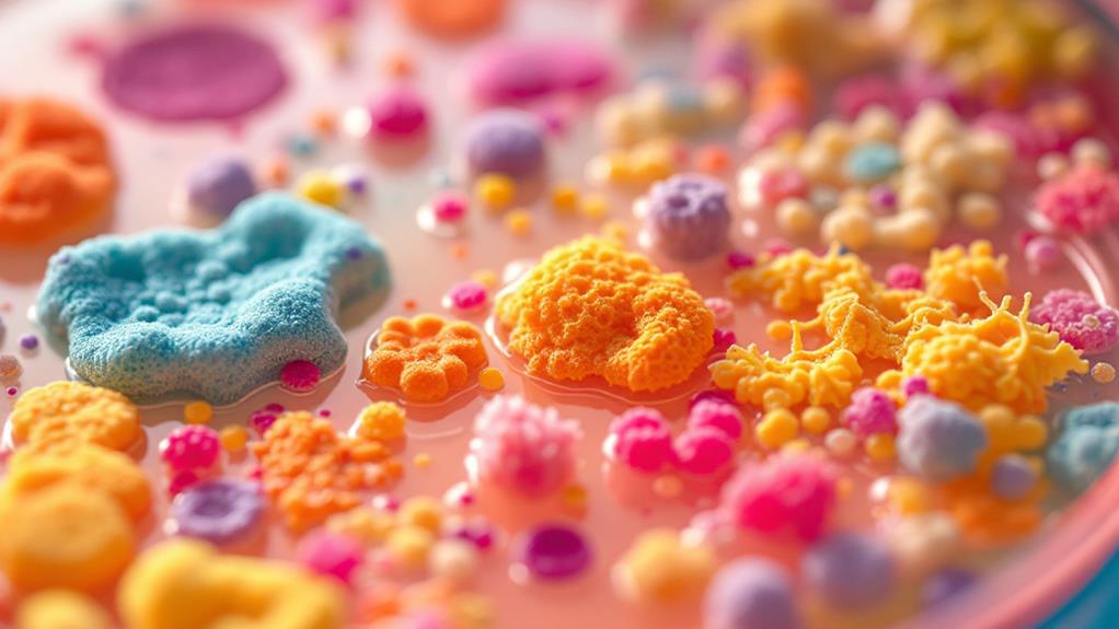
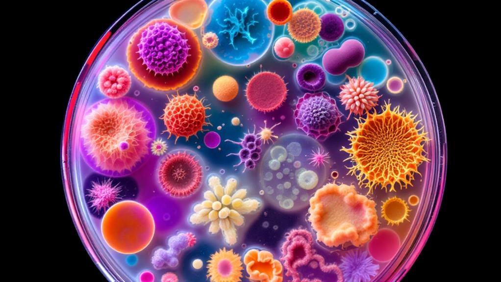
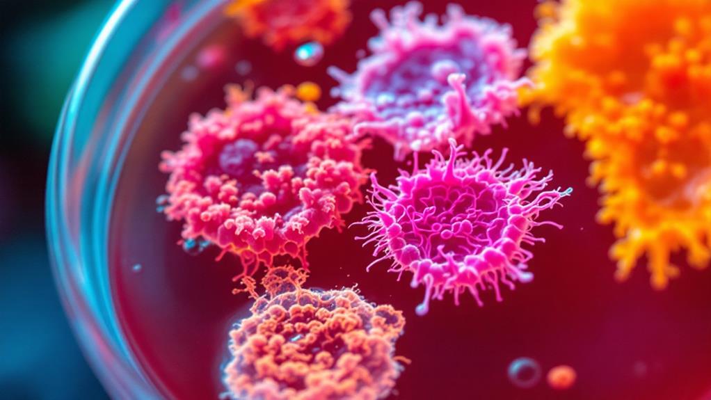
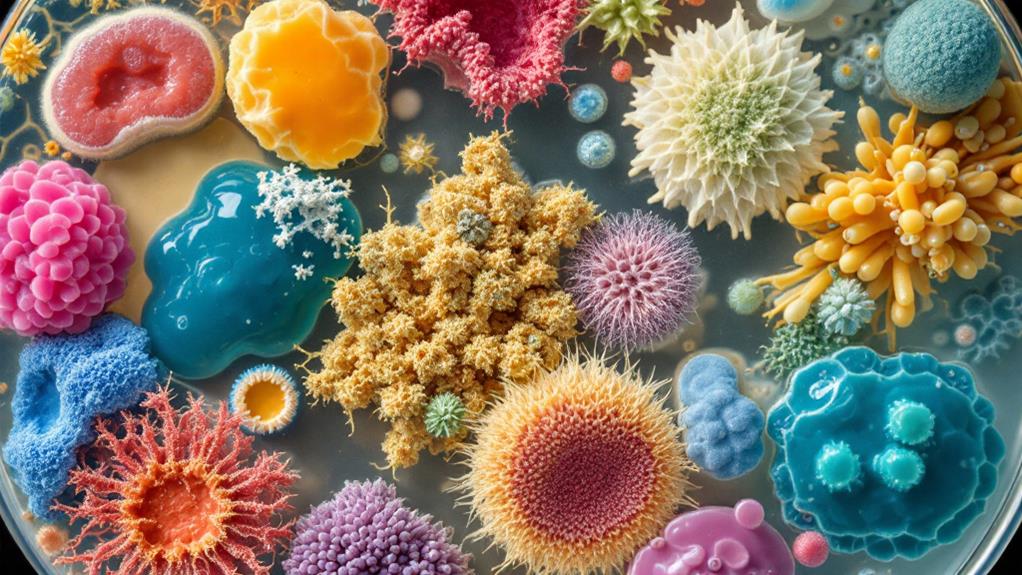
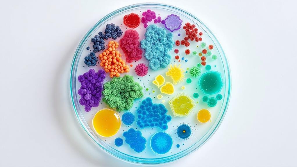
Bacterial colony morphology types provide essential clues about microbial species and their growth patterns. You’ll observe various characteristics, including form and shape (circular, irregular, rhizoid), size and elevation (pinpoint to large, flat to umbonate), margin features (entire, undulate, lobate), color and pigmentation, and surface texture (smooth, rough, mucoid). Optical properties like opacity, luster, and fluorescence offer additional insights. Environmental factors such as temperature, pH, nutrient availability, and oxygen levels greatly influence colony appearance. By understanding these morphological traits, you’ll be better equipped to identify and differentiate bacterial species. Exploring these features further can reveal a wealth of microbiological knowledge.
Form and Shape
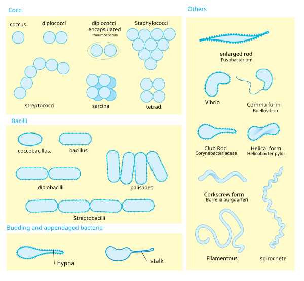
The form and shape of bacterial colonies are key characteristics used to identify and classify different bacterial species. When you’re examining bacterial cultures, you’ll notice that colonies can take on various forms. The most common shapes you’ll encounter are circular, irregular, and rhizoid.
Circular colonies have smooth, round edges and are typically symmetrical. You’ll often see these in species like Staphylococcus aureus. Irregular colonies, as the name suggests, have uneven edges and lack a defined shape. Bacillus species frequently display this morphology. Rhizoid colonies are characterized by root-like extensions that spread out from the center, resembling a branching tree. You’ll find this pattern in certain Bacillus species, like Bacillus mycoides.
Other forms you might observe include punctiform (tiny, dot-like colonies), filamentous (thin, thread-like growth), and spindle-shaped (elongated with tapered ends). Each of these forms can provide clues about the bacterial species you’re dealing with.
When examining colony shape, pay attention to the elevation as well. Colonies can be flat, raised, convex (domed), or umbonate (raised with a central knob). The margin of the colony is another important feature. It can be entire (smooth), undulate (wavy), lobate (lobed), or filamentous (hair-like projections).
Remember that environmental factors, such as nutrient availability and incubation conditions, can influence colony morphology. Always consider these variables when interpreting your observations. By carefully noting the form and shape of bacterial colonies, you’ll gain valuable insights into the organisms you’re studying.
Size and Elevation
While form and shape provide valuable information about bacterial colonies, size and elevation offer additional important clues for identification. When examining bacterial colonies, you’ll notice variations in size ranging from pinpoint to several millimeters in diameter. Size can be influenced by factors such as growth rate, nutrient availability, and incubation time.
You’ll typically categorize colony sizes as:
- Pinpoint: Less than 1mm in diameter
- Small: 1-2mm in diameter
- Medium: 2-4mm in diameter
- Large: More than 4mm in diameter
It’s important to note that colony size can change over time, so you should always record observations at a specific point in incubation.
Elevation refers to the height of the colony above the agar surface. You’ll observe various elevation types, including:
- Flat: No discernible raise above the agar
- Raised: Slightly elevated above the surface
- Convex: Dome-shaped with a curved surface
- Pulvinate: Cushion-shaped with a flattened top
- Umbonate: Raised with a central knob
- Crateriform: Sunken center with elevated edges
When examining elevation, you’ll want to view the colonies from the side to accurately assess their height and profile. Some bacteria produce characteristic elevations, which can aid in identification. For example, Staphylococcus aureus typically forms convex colonies, while Bacillus cereus often produces flat colonies with irregular edges.
Margin Characteristics
Bacterial colonies’ margins provide vital information about their characteristics and can aid in identification. When examining bacterial colonies, you’ll notice various margin types that can help you distinguish between different species or strains. These margin characteristics are significant for microbiologists and researchers in understanding bacterial growth patterns and identifying specific organisms.
The most common margin types you’ll encounter include:
- Entire: A smooth, even edge with no irregularities.
- Undulate: A wavy edge that resembles gentle curves.
- Lobate: Large, rounded projections along the edge.
- Erose: An irregular, eroded-looking edge.
- Filamentous: Hair-like projections extending from the margin.
- Rhizoid: Root-like extensions branching out from the edge.
To observe these margin characteristics, you’ll need to use a stereomicroscope or a magnifying glass. It’s essential to recognize that some bacteria may exhibit different margin types depending on their growth conditions or age.
When you’re identifying bacteria based on colony morphology, you should consider margin characteristics alongside other features like size, elevation, and pigmentation. For example, Bacillus cereus often displays undulate margins, while Serratia marcescens typically has entire margins.
You’ll find that margin characteristics can change as colonies age or are exposed to different environmental conditions. It’s best to observe colonies at various growth stages to get an all-encompassing understanding of their morphology.
Color and Pigmentation
Color and pigmentation play an essential role in bacterial colony identification. When you’re examining bacterial colonies, you’ll notice a wide range of colors and pigments that can provide valuable information about the species and their characteristics.
You’ll commonly encounter white or off-white colonies, which are typical for many bacterial species. However, some bacteria produce distinctive pigments that can help you quickly identify them. For instance, Serratia marcescens forms bright red colonies due to its prodigiosin pigment, while Pseudomonas aeruginosa often displays a blue-green color from pyocyanin production.
Yellow pigments are also prevalent, such as those produced by Staphylococcus aureus or some Micrococcus species. You might observe golden-yellow, lemon-yellow, or even orange hues. Some Bacillus species can create colonies with a pinkish tinge, while certain Corynebacterium strains may appear grayish.
It’s important to note that pigmentation can vary depending on growth conditions and media composition. You should always consider these factors when evaluating colony color. Additionally, some bacteria may produce diffusible pigments that can change the color of the surrounding medium.
As you gain experience, you’ll find that color and pigmentation become valuable tools in your bacterial identification arsenal. However, remember that while color can provide initial clues, it shouldn’t be used as the sole criterion for identification. Always combine your observations of color with other morphological characteristics and biochemical tests for accurate bacterial identification.
Surface Texture
In addition to color, the surface texture of bacterial colonies provides essential information for identification. When examining bacterial colonies, you’ll notice various surface characteristics that can help you distinguish between different species.
One of the primary textures you’ll encounter is smooth. Smooth colonies have a uniform, glossy appearance and feel slick to the touch. They’re common in many bacterial species, including Escherichia coli and Staphylococcus aureus.
You might also observe rough colonies, which have an uneven, granular surface. These colonies often appear dull and feel coarse when touched. Mycobacterium tuberculosis is a well-known example of a bacterium that forms rough colonies.
Some bacteria produce mucoid colonies, which are characterized by a slimy or gooey texture. These colonies are often raised and have a shiny, wet appearance. Klebsiella pneumoniae typically forms mucoid colonies due to its capsule production.
You’ll find that certain bacteria form colonies with a dry, powdery texture. These are often described as pulvinate or powdery. Bacillus anthracis, the causative agent of anthrax, is known for its dry, powdery colonies.
When examining colonies, you may come across those with a wrinkled or folded surface. These are called rugose colonies and are often associated with bacterial species that produce extracellular polysaccharides. Vibrio cholerae can form rugose colonies under certain conditions.
Optical Properties
When examining bacterial colonies, you’ll notice that their optical properties can provide valuable insights into their characteristics. These properties refer to how the colonies interact with light, which can help you differentiate between various bacterial species and strains.
One of the primary optical properties you’ll observe is opacity. Some colonies appear transparent, allowing light to pass through them easily. Others are translucent, partially allowing light transmission, while opaque colonies block light completely. The degree of opacity can indicate the density of bacterial cells or the presence of certain pigments.
Color is another vital optical property you’ll encounter. Many bacterial colonies exhibit distinct hues, ranging from white and cream to vibrant yellows, reds, or even purples. These colors often result from pigments produced by the bacteria, which can serve various functions such as protection from UV radiation or antibiotic production.
You’ll also want to pay attention to the colony’s luster or shininess. Some colonies appear glossy or shiny, while others have a dull or matte appearance. This property can be influenced by the production of extracellular substances or the arrangement of bacterial cells within the colony.
Fluorescence is an interesting optical property you might observe in certain bacterial species. Under UV light, these colonies emit a distinctive glow, which can be useful for identification purposes.
Lastly, you’ll want to note any iridescence or color changes that occur when viewing the colony from different angles. This phenomenon, known as structural color, can result from the unique arrangement of bacterial cells or the production of specific compounds.
Environmental Influences
Environmental factors frequently play an essential role in shaping bacterial colony morphology. As you observe bacterial colonies, you’ll notice that their appearance can change dramatically based on the conditions in which they’re grown. Temperature, pH, nutrient availability, and oxygen levels are just a few of the factors that can influence colony characteristics.
Temperature affects bacterial growth rates and metabolism, which in turn impacts colony size and shape. Some bacteria thrive in cooler environments, while others prefer warmer conditions. You’ll find that colonies grown at their ideal temperature tend to be larger and more well-defined.
The pH of the growth medium can alter colony texture and pigmentation. Acidic or alkaline conditions may stress bacteria, leading to smaller, more compact colonies or unusual coloration. You’ll need to take into account pH when interpreting colony morphology, as it can greatly impact the appearance of certain species.
Nutrient availability directly affects colony size and density. In nutrient-rich media, you’ll observe larger, more robust colonies. Conversely, nutrient-poor conditions result in smaller, slower-growing colonies. The type of nutrients available can also influence pigment production and overall colony appearance.
Oxygen levels play a critical role in determining colony morphology for aerobic and anaerobic bacteria. You’ll notice that aerobic bacteria form larger, more spread-out colonies when grown in oxygen-rich environments, while anaerobic bacteria may exhibit stunted growth or fail to form colonies altogether in the presence of oxygen.

Erzsebet Frey (Eli Frey) is an ecologist and online entrepreneur with a Master of Science in Ecology from the University of Belgrade. Originally from Serbia, she has lived in Sri Lanka since 2017. Eli has worked internationally in countries like Oman, Brazil, Germany, and Sri Lanka. In 2018, she expanded into SEO and blogging, completing courses from UC Davis and Edinburgh. Eli has founded multiple websites focused on biology, ecology, environmental science, sustainable and simple living, and outdoor activities. She enjoys creating nature and simple living videos on YouTube and participates in speleology, diving, and hiking.